Skin Barrier · Foundations · Approximately 7 minutes
Understanding Our Skin — Anatomy & Function
The science behind the organ we care for every day.
By Anna Olechowska, MPharm, MRPharmS, Independent Prescriber
Our skin is living, responsive tissue. It helps protect us from our surroundings, senses touch and temperature, supports immune defence and helps regulate body temperature. The surface we see is just one part of a much larger system. Understanding what sits beneath it gives us a useful starting point for making sense of skincare claims. [1,2]
Three connected regions, different responsibilities
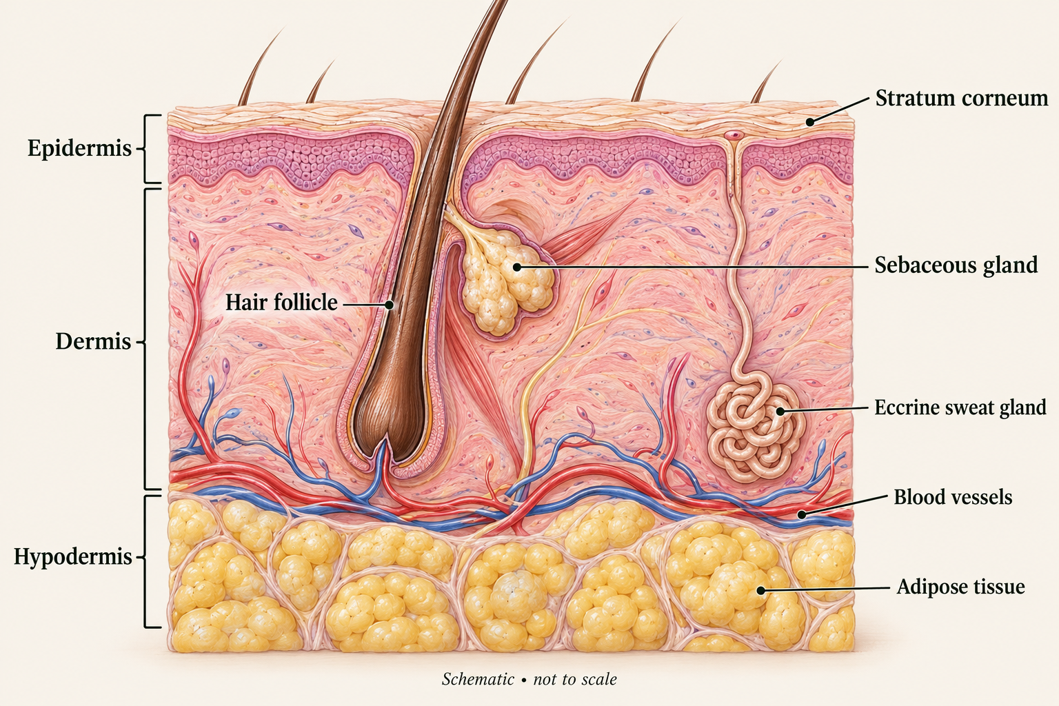
We often describe our skin using three regions: the epidermis, dermis and hypodermis. Strictly speaking, skin itself comprises the epidermis and dermis; the hypodermis is the closely associated subcutaneous tissue beneath them. Showing all three helps us understand how the surface connects to the tissues below. [1]
The epidermis is the outer cellular covering. The dermis is the connective tissue beneath it, providing mechanical support and housing vessels, nerves and many skin appendages. The hypodermis contains fat and connective tissue that cushion and insulate us. Their thickness and arrangement vary across our body. [1,2]
Our epidermis: a living tissue with a specialised surface
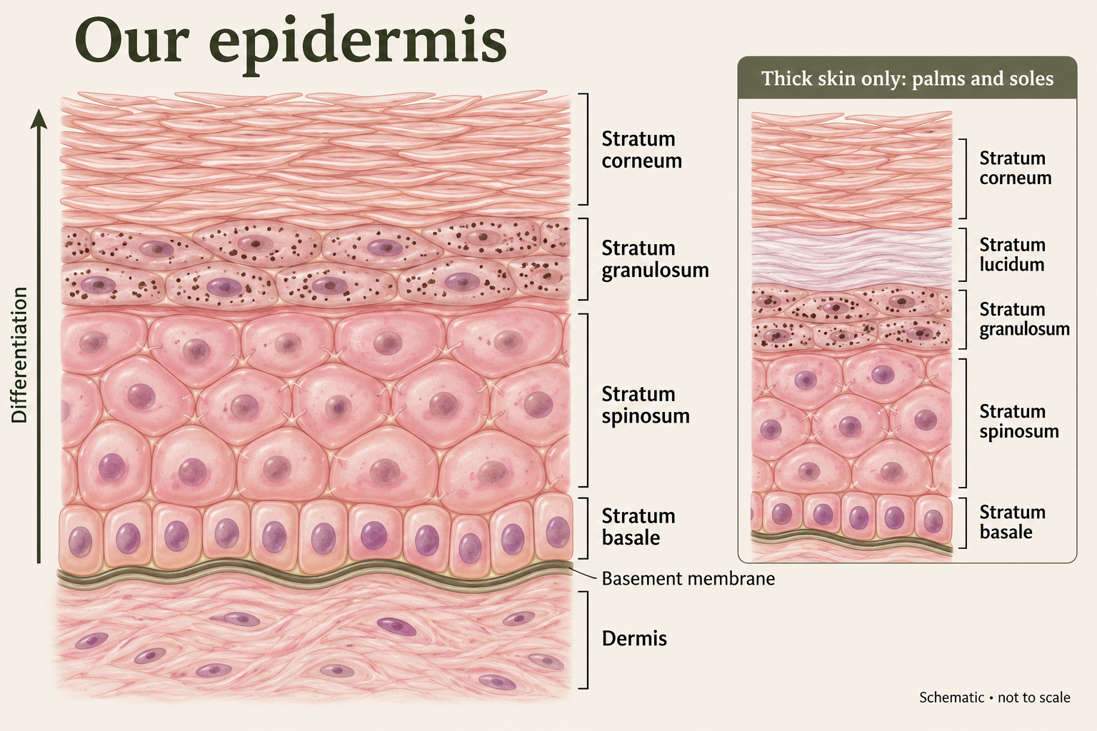
Most epidermal cells are keratinocytes. They produce keratin proteins and change their structure as they mature towards the surface. The epidermis has no blood vessels of its own: oxygen and nutrients reach its living cells by diffusion from the underlying dermal circulation. A specialised basement membrane anchors and separates the epidermis from the dermis. [2]
The five epidermal strata, from deep to superficial
1. Stratum basale — the basal layer. This deepest layer is usually one cell thick. It contains proliferating keratinocytes, including stem and progenitor cells that sustain epidermal renewal. Not every cell divides at the same moment. [2]
2. Stratum spinosum — the spinous layer. Keratinocytes become more differentiated and remain connected by strong junctions called desmosomes. The familiar “spiny” appearance seen in prepared tissue sections reflects these connections and the way cells shrink during processing. [2]
3. Stratum granulosum — the granular layer. Cells flatten and develop prominent keratohyalin granules. They undergo coordinated changes that prepare them to become the specialised cells of the outer surface. [2,3]
4. Stratum lucidum — the clear layer. This thin, translucent layer is identifiable in thick skin on our palms and soles. It lies between the granular and cornified layers. It is not a normal distinct layer of facial skin. In anatomy, “thick skin” describes this particular tissue organisation, rather than simply skin that feels thick. [1]
5. Stratum corneum — the cornified layer. The outermost layer consists of flattened corneocytes: terminally differentiated keratinocytes that have lost their nuclei and organelles. These cells are essential components of our principal permeability barrier, helping limit water loss and entry of external substances. They are functional protective structures, even though they are no longer living cells. [3]
Most of our skin has four clearly recognised epidermal strata; thick skin has five. [1]
The other cells that help our epidermis work
Keratinocytes do more than provide structure. They also send signals involved in immune responses and communicate with neighbouring cells. [4]
Melanocytes, found in the basal epidermis, produce melanin and transfer pigment-containing structures to keratinocytes. Melanin contributes to our skin colour and helps protect cellular DNA against ultraviolet radiation. It does not provide complete protection from sun damage. [2,5]
Langerhans cells are immune cells found mainly among keratinocytes in the spinous layer. They sample material in their surroundings and help coordinate immune responses. [5,6]
Merkel cells lie in the basal layer and form complexes with sensory nerve endings. They contribute to fine touch perception. Our skin’s sensory function therefore begins very close to its surface. [5]
Our dermis: strength, elasticity and support
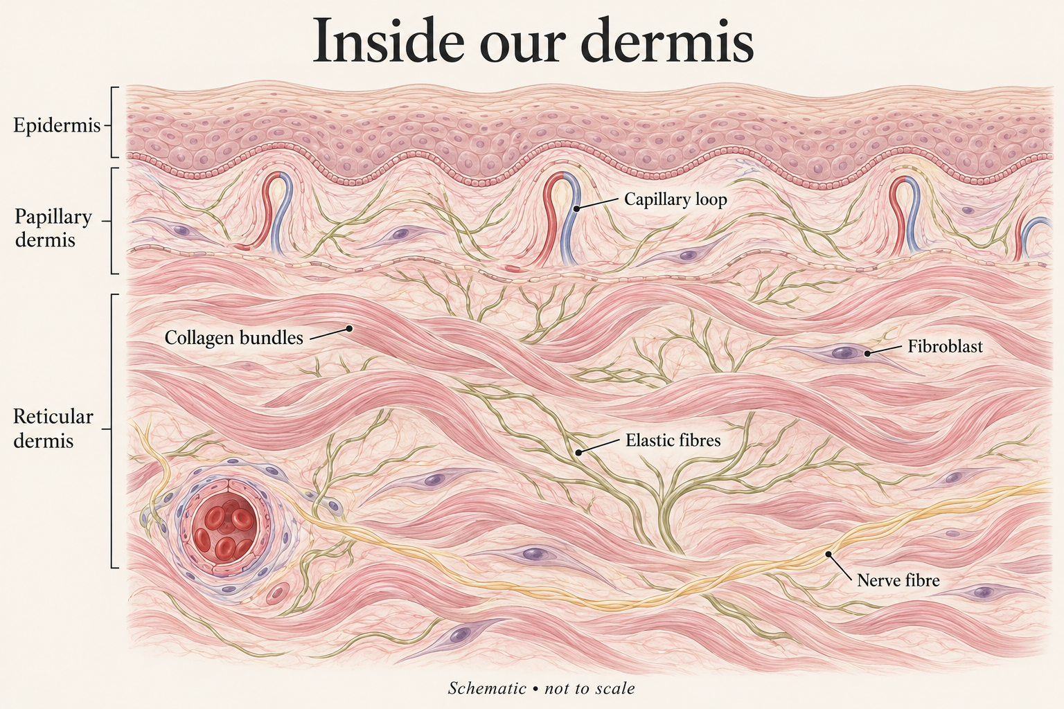
The dermis is a living connective tissue network. Its upper papillary region has finer connective tissue and small vessels close to the epidermis. Its deeper reticular region contains denser, interwoven bundles of collagen. These regions blend into one another. [7]
Collagen provides tensile strength: it helps our skin resist being pulled apart. Elastic fibres, which contain elastin, help it stretch and recoil. Fibroblasts produce and maintain many components of the surrounding extracellular matrix, the material between cells. This matrix also includes molecules that bind water and help organise the tissue. [7,8]
Our dermis contains blood and lymphatic vessels, immune cells and nerves. Hair follicles and their associated sebaceous glands occupy many areas of skin; sebaceous glands release sebum into follicles. Eccrine sweat glands have coiled secretory portions deep in the skin and ducts that open onto the surface. Their sweating helps us release heat. Distribution varies: palms and soles have eccrine glands but no hair follicles or sebaceous glands. [1,7]
Where skin ageing fits into the picture
With ageing, changes in the dermal matrix and fibroblast activity affect the skin’s mechanical properties. Repeated ultraviolet exposure adds photoageing, including changes to collagen and elastic tissue. These processes help explain why surface appearance reflects more than what is happening in the epidermis. [8,9]
Knowing that collagen sits in the dermis does not tell us whether a particular product can change it. An ingredient must be delivered appropriately, and a claim needs evidence for the formulation, use and outcome being discussed. A laboratory finding is a starting point, rather than proof of a visible benefit from a finished skincare product.
Our hypodermis: cushioning beneath the skin
The hypodermis contains adipose tissue arranged in lobules separated by connective tissue. It provides cushioning, insulation and an energy store, while helping connect the skin to deeper structures. Larger vessels and nerves travel through this region. Its thickness varies with body site and between individuals. [1,2]
How our epidermis renews
Renewal is a sequence, rather than a single event. Basal keratinocytes divide; some of their descendants leave the basal layer and differentiate as they move upwards. Through cornification, they become the specialised corneocytes of the stratum corneum. Finally, surface cells detach through desquamation. Cell production, differentiation and shedding are related, but different processes. [3,4]
Our skin does not renew according to one universal “28-day” clock. Estimates depend on what is measured, and renewal varies with body site, age and skin health. Normal shedding is often invisible. Visible peeling does not, by itself, demonstrate that a product is working well.
Science of Beauty Rx explains: what this means for skincare
Our surface deserves care. The stratum corneum is a protective structure, so removing more surface cells is not automatically better. Exfoliation is not a requirement for everyone.
Deeper is not automatically better. Many useful skincare effects occur at or near the surface. The location of a biological target does not establish how far a topical ingredient will penetrate.
Claims need context. When a product promises to “stimulate collagen” or “speed renewal”, ask what was studied, in whom, and whether the result was measured in people using the finished product.
Our skin is a connected organ. Its appearance reflects interactions between surface cells, pigment, immune signalling, connective tissue and the tissues beneath it. A thoughtful routine starts with understanding those functions, rather than treating every visible feature as something to correct.
Next in this series: Understanding Our Skin Barrier. We will look more closely at ceramides, water loss, natural moisturising factor, pH and barrier repair in Article 2.
Explore the education library →
Educational information; individual skin concerns may need personalised assessment.
References
1. OpenStax. Anatomy and Physiology 2e. Section 5.1: Layers of the Skin. Read source ↗
2. StatPearls. Anatomy, Skin (Integument), Epidermis. NCBI Bookshelf. Read source ↗
3. Segre JA. Epidermal barrier formation and recovery in skin disorders. Journal of Clinical Investigation. 2006;116:1150–1158. Read source ↗
4. Cytokinocytes: the diverse contribution of keratinocytes to immune responses in skin. JCI Insight. 2021. Read source ↗
5. Nothing but skin and bone. Journal of Clinical Investigation. 2006. Read source ↗
6. CD1a and langerin: acting as more than Langerhans cell markers. Journal of Clinical Investigation. 2004. Read source ↗
7. StatPearls. Histology, Dermis. NCBI Bookshelf. Read source ↗
8. Quan T, Fisher GJ. Extracellular matrix regulation of fibroblast function: redefining our perspective on skin aging. Cell and Tissue Research. 2018. Read source ↗
9. Fisher GJ et al. Mechanisms of photoaging and chronological skin aging. Archives of Dermatology. 2002;138:1462–1470. Read source ↗